
Known in medical literature as "Macular Degeneration," yellow spot disease is a condition where the macula (yellow spot) region, located at the back of the eye and responsible for sharp, clear, and detailed vision, becomes damaged over time.
Learn More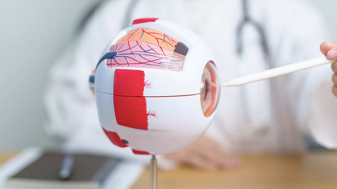
Diabetic retinopathy is a condition where the capillaries of the retina—the inner layer at the back of the eye that transmits visual signals to the brain—are damaged due to blood sugar remaining high and uncontrolled for a long time.
Learn More
The retina (nerve layer), which covers the back inner wall of our eye like wallpaper, consists of millions of nerve cells that perceive light from the outside and transmit it to the brain via optic nerves.
Learn More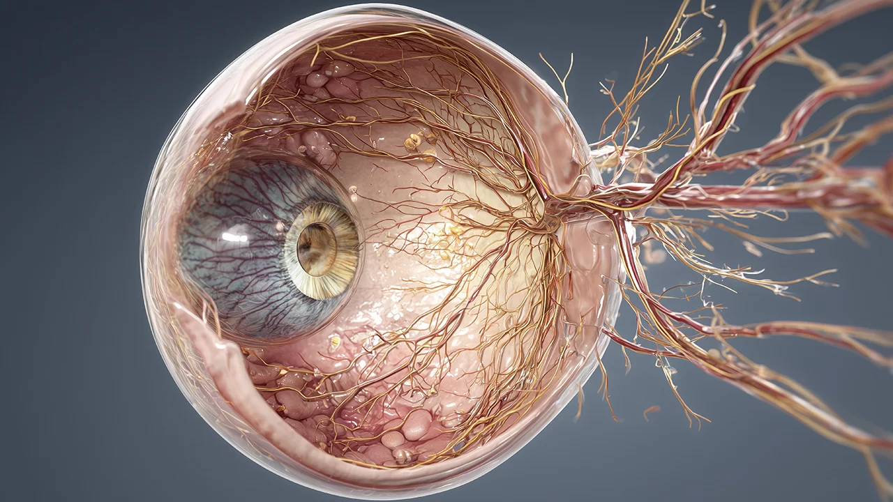
Just as our heart or brain needs an uninterrupted blood flow to function, the nerve layer of our eye (retina) similarly requires rich oxygen and nutrient support. The retina is nourished by a main artery (artery) coming from the center and sends back used, deoxygenated blood through a main vein (vein) also exiting from the center.
Learn More
In a normal pregnancy process, vascular development in the baby's eye begins in the womb at approximately the 16th week and continues until birth (around the 40th week), reaching the far edges of the retina (the nerve layer of the eye). However, when a baby is born prematurely, this normal vascular development process is interrupted.
Learn More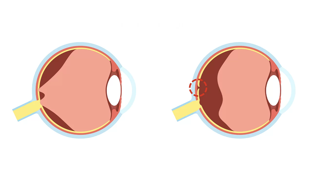
The nerve layer covering the back inner wall of our eye, consisting of light-sensitive nerve cells, is called the retina. The millimetric area located at the very center of the retina, approximately the size of a pinhead, is called the macula (yellow spot).
Learn More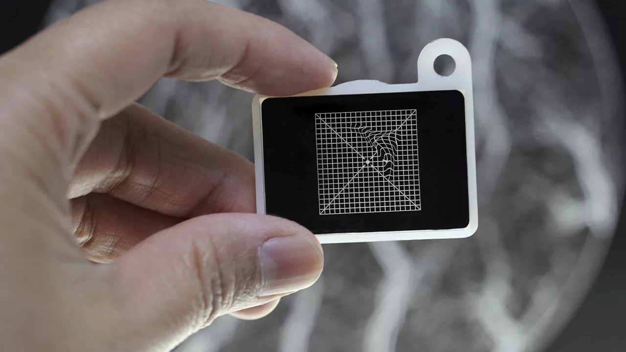
Also known in medical literature as "Macular Pucker," "Cellophane Maculopathy," or "Premacular Fibroplasia," Epiretinal Membrane (ERM) is a thin, semi-transparent, and membrane-like scar tissue that forms directly over the retina—the innermost layer of the eye—and specifically the macula (yellow spot) region responsible for sharp, detailed vision.
Learn More
The nerve layer covering the inner surface of our eye and initiating the vision process is called the retina. The most critical region of the retina is the macula (yellow spot), our sharp vision center responsible for functions such as reading, recognizing faces, and clearly distinguishing colors.
Learn More
Retinitis Pigmentosa (RP) is not a single disease but a general name for a broad group of hereditary retinal dystrophies that cause the light-sensitive cells of the retina to degenerate slowly and irreversibly.
Learn More
Known in medical literature as "Juvenile Macular Degeneration" or "Fundus Flavimaculatus," Stargardt Disease is a hereditary condition that directly targets the "macula" (yellow spot)—the center of the retina responsible for sharp, clear vision.
Learn More
First described in 1869 by Dr. Theodor Leber, Leber Congenital Amaurosis (LCA) is one of the most common genetic causes of congenital blindness in childhood. It is a group of severe and early-onset hereditary retinal dystrophies that cause the retina (the light-sensitive nerve layer at the back of the eye) to fail in its function from birth.
Learn More
In medicine, "congenital" means "present at birth," and "dystrophy" refers to a tissue or organ (in this case, the retina) failing to complete its normal development, becoming structurally impaired, and losing function due to genetic errors.
Learn More
The nerve layer covering the back inner wall of our eye is called the retina, and the millimetric area located at the exact center of this layer, responsible for sharp, colored, and detailed vision, is called the "macula" (yellow spot). Reading books, driving, threading a needle, or recognizing a person's face depends entirely on the healthy functioning of the macula.
Learn More
To maintain the vitality and spherical shape of the eyeball and to nourish avascular tissues like the cornea and lens, a special fluid called "aqueous humor" is constantly produced inside the eye. In a healthy eye, this fluid leaves through microscopic channels called the "trabecular meshwork" in the anterior chamber and enters the bloodstream.
Learn More
Toxic Optic Neuropathy occurs when poisonous chemicals, heavy metals, or certain powerful drugs reach the eye via the bloodstream, poisoning and killing these vital nerve fibers at a cellular level. The most severe, rapid, and unfortunately, most lethal subtype of this condition is Methyl Alcohol (Methanol) Intoxication.
Learn More
The optic nerve is a massive network of approximately 1.2 million fine nerve fibers that collects light and color signals formed in the retina (the back layer of the eye) and transmits them to the visual center (occipital cortex) at the back of the brain.
Learn More
Stem cells are the fundamental building blocks—the "master" cells—of the human body. They form the origin of all other specialized cells (such as heart muscle, brain nerves, or retinal cells).
Learn More
Exosomes are, in the simplest terms, nano-sized (one-millionth of a millimeter) "cargo packages" or "messenger vesicles" that cells use to communicate, "talk," and help one another.
Learn More
PRP (Platelet-Rich Plasma) is a natural, safe, and highly effective cellular regeneration treatment obtained entirely using the patient's own blood. Our blood contains specialized cells called "platelets" that are the first to arrive at any site of injury or tissue damage to initiate the healing process.
Learn More
In hereditary retinal diseases (such as advanced-stage Retinitis Pigmentosa), the primary light-sensing cells, the photoreceptors (rods and cones), completely die over time. When photoreceptors are lost, standard gene therapies (such as Luxturna) become ineffective because there are no living cells left for the therapy to repair.
Learn More
In the nucleus of every cell in the human body lies a manual that determines how that cell works, which proteins it will produce, and how it will survive: the DNA (genetic code). The light-sensitive cells in the retina, which initiate the process of vision, also operate according to these genetic instructions.
Learn More
Ocuvision therapy, referred to in medical literature as "Transcorneal Electrical Stimulation (TES)," is an innovative, non-surgical, and highly reliable treatment method based on the principle of sending specific, very low-dose, controlled electrical currents to the outer surface of the eye (cornea) using the specially developed OkuStim device.
Learn More
Referred to in medical literature as "Photobiomodulation (PBM)," Valeda Light Therapy is the process of using light at very specific and carefully selected wavelengths (yellow, red, and near-infrared) to heal damaged cells in the posterior layer of the eye (retina).
Learn More
Based on the principles of "Repetitive Electromagnetic Stimulation (rEMS)" or "Transcranial Magnetic Stimulation (rTMS)" in medical literature, Magnovision therapy is the process of stimulating the retinal cells at the back of the eye and the optic (visual) nerve fibers leading to the brain using powerful but short-term external magnetic fields.
Learn More
When the macular tissue responsible for central vision dies due to disease or trauma, it may not always be possible to fully revitalize those cells with modern medicine. The patient sees only a dark or blurred area when looking directly ahead.
Learn More
The human eye functions similarly to a perfect camera. Right behind the iris (the colored part of our eye), there is an entirely transparent, flexible, and crystalline "natural intraocular lens" that refracts incoming light rays and focuses them onto the retina (the visual center).
Learn More
In a healthy eye, the "natural intraocular lens" located right behind the iris refracts incoming light rays so they fall clearly onto the retina. To perform this function, the lens must be completely transparent and clear, like clean glass.
Learn More
In a healthy eye, the natural intraocular lens located right behind the iris refracts light and focuses it onto the retina. This lens is protected within a transparent and highly sensitive capsule (membrane).
Learn More
In a standard cataract surgery (Primary Lens Application), the opaque natural lens is cleared using ultrasonic waves, but the outer membrane (capsule) is left in place. The artificial lens is then tucked into this natural "pocket."
Learn More
In youth, the natural lens inside our eye acts like an autofocus motor. When we look at distance, near, or intermediate ranges, it instantly changes shape (flexes) to focus the image clearly onto the retina.
Learn More
During cataract surgery (the PHACO method), your natural lens is removed, but the transparent "capsule" that surrounds it is intentionally left in place. This clear back wall acts like a thin membrane, providing vital support to keep your new Artificial Intraocular Lens (IOL) perfectly in its housing.
Learn More
Our eyes are a unique pair of cameras that work in perfect synchronization and harmony, allowing us to perceive the world with depth, color, and reality. For the brain to create a single, clear, three-dimensional image, it is vital that both cameras focus on the same target. However, Strabismus (Eye Misalignment), which occurs due to an imbalance between the muscles that move the eyes, disrupts this harmony and deeply affects patients' quality of life both functionally and aesthetically.
Learn More
Lazy eye is a condition where the vision capacity of an eye fails to reach normal levels (100%), even though there is no anatomical or structural damage (tears, scars, tumors, etc.) to the eyeball itself. This problem actually occurs in the brain, not the eye.
Learn More
The eye, in its basic principle, is a perfect optical system. Incoming light passes through the transparent outer layer (cornea) and is refracted by the internal lens to focus exactly on the visual center at the back of the eye (retina). When light falls precisely on the retina, a sharp image is sent to the brain.
Learn More
Just as we learn to use a walker and undergo physical therapy to walk again after a leg injury, the brain must learn to "see again" and adapt to new conditions following cellular damage in the eye. Low vision exercises are a highly specific neuro-cognitive form of physical therapy adapted for the eye and brain.
Learn More
Stem cells are the fundamental building blocks—the "master" cells—of the human body. They form the origin of all other specialized cells (such as heart muscle, brain nerves, or retinal cells).
Learn More
Exosomes are, in the simplest terms, nano-sized (one-millionth of a millimeter) "cargo packages" or "messenger vesicles" that cells use to communicate, "talk," and help one another.
Learn More
When tissues in our body sense a lack of oxygen, they release a protein called Vascular Endothelial Growth Factor (VEGF) to create new blood vessels. While usually beneficial, this protein can go out of control in certain eye diseases.
Learn More
While many people associate "eye laser" only with getting rid of glasses (Excimer Laser/LASIK), therapeutic lasers are vital tools that often prevent permanent blindness.
Learn More
The interior of the eyeball is not empty; it is filled with a transparent, egg-white-like gel called the Vitreous. This gel is firmly attached to the retina at the back of the eye. Due to aging, trauma, or diabetes, this gel can lose its structure, fill with blood, or shrink and pull on the retina, causing a tear.
Learn More
The fundamental logic of treating retinal detachment is to reattach the displaced nerve layer (retina) to the outer wall of the eye (sclera). While a Vitrectomy does this by entering the eye and applying pressure from the inside out (using gas or silicone), Scleral Buckling works with the opposite mechanism: from the outside in.
Learn More
If we compare the eye to a high-resolution camera, the most critical component is the macula (yellow spot) located at its very center. This tiny area, only a few millimeters in diameter, is responsible for our "HD" vision—allowing us to recognize faces, read books, distinguish vibrant colors, and drive safely.
Learn More
OCT technology functions similarly to ultrasonography (USG), but instead of sound waves, it uses low-energy, specialized laser light (near-infrared, around 840 nm).
Learn More
Directly beneath the retina lies a vital layer called the Retinal Pigment Epithelium (RPE), which acts as a "waste processing plant" for the eye. As we age or face genetic diseases, this plant becomes fatigued, and a toxic aging pigment called Lipofuscin begins to accumulate within the cells.
Learn More
Traditional fundus cameras and standard examination methods can only capture a narrow 30 to 45-degree area at the center of the retina at one time. This represents only about 15% of the total retinal surface.
Learn More
Vision is more than just an optical process; it is a magnificent "bioelectrical" miracle. Light entering the eye is instantly converted into electrical signals by millions of microscopic cells and transmitted to the brain's visual center via a biological cable network called the optic nerve.
Learn More
The visual field is the entire spatial area you can perceive (up, down, left, and right) while keeping your head and eyes fixed on a single point. A healthy human eye can scan approximately 160 degrees horizontally and 120 degrees vertically.
Learn More
In the field of eye health, the ability to read letters on a chart or having an anatomically sound eye structure does not always equate to high-quality vision. Often, the subtle "working capacity" (function) of the cells—which standard exams might miss—is the true measure of how we perceive the world.
Learn More