Eye health is one of the most important factors determining our quality of life. Macular Degeneration (Age-Related Macular Degeneration), which can occur in our eyes with advancing age and directly affects central vision, is a condition that can be kept under control through early diagnosis and correct treatment methods.
What is Macular Degeneration (Age-Related Macular Degeneration)?
Known in medical literature as "Macular Degeneration," yellow spot disease is a condition where the macula (yellow spot) region, located at the back of the eye and responsible for sharp, clear, and detailed vision, becomes damaged over time. The macula is of critical importance for daily vital activities such as reading, driving, recognizing faces, and discerning fine details. The loss of function in the cells of this region results in blurred central vision, dark spots, or distortions. The disease usually does not affect peripheral (side) vision; therefore, patients do not go completely blind, but they cannot clearly see the point they are focusing on.
Macular degeneration is clinically divided into two main types:
1. Dry Type Macular Degeneration (Dry AMD)
This is the most common form, accounting for approximately 85-90% of all macular degeneration patients. It begins with the formation of yellow-colored protein and fat deposits called "drusen" in the tissues under the macula. Over time, these deposits cause the light-sensitive cells in the macula to thin and dry out. Vision loss generally progresses slowly and gradually. The dry type carries the risk of transforming into the more serious wet type over time.
2. Wet Type Macular Degeneration (Wet AMD)
Although less common, the wet type is responsible for the majority of serious vision loss associated with macular degeneration. It can occur as the dry type progresses or may appear suddenly. It is characterized by the formation of abnormal, weak, and fragile new blood vessels under the retina. These new vessels leak easily, leading to the accumulation of blood and fluid under the macula. This fluid accumulation rapidly disrupts the structure of the macula and causes sudden, severe loss of central vision.
What are the Causes and Risk Factors of the Disease?
Although the exact cause of macular degeneration is not fully known, it is thought to occur as a result of a complex interaction of genetic and environmental factors. The most significant risk factors include:
- Age: The greatest risk factor. The incidence generally increases in individuals aged 50 and over.
- Genetic Predisposition: People with a family history of macular degeneration have a much higher risk of developing this disease.
- Smoking: Smoking or being a passive smoker significantly increases the risk of disease development and the rate of progression.
- Obesity and Poor Nutrition: A diet poor in antioxidants, vitamins, and minerals, combined with excess weight, negatively affects macular health.
- Cardiovascular Diseases: High blood pressure, high cholesterol, and diseases of the cardiovascular system also affect the vascular structure of the eye, raising the risk.
- Sunlight: Exposing the eyes to UV rays for long periods without protection can accelerate cellular damage.
What are the Symptoms of Macular Degeneration?
In the early stages of the disease, especially in the dry type, no obvious symptoms may be seen. However, as the disease progresses, the following symptoms appear:
- Blurring or shadowing in central vision.
- Straight lines (e.g., door frames or tile lines) appearing wavy, broken, or bent (Metamorphopsia).
- Needing more light when reading or doing fine work.
- Difficulty recognizing people's faces.
- The appearance of a dark or blank spot (scotoma) right in the center of the visual field.
- Colors losing their vibrancy and appearing faded.
How is it Diagnosed? Our Advanced Diagnostic Methods
When our health tourism patients apply to our clinic, a comprehensive eye examination is performed with our world-class technological infrastructure. The diagnostic methods applied by Ophthalmology Specialist Ayşe Öner are as follows:
- Detailed Fundus Examination: The pupils are dilated with drops, and the condition of the retina and macula is examined in detail with special lenses.
- Optical Coherence Tomography (OCT): A highly advanced device that takes microscopic-level, radiation-free 3D cross-sectional images of the eye.
- Fluorescein Angiography (FFA): A special, harmless dye is administered through an arm vein, and photos of the vessels at the back of the eye are taken.
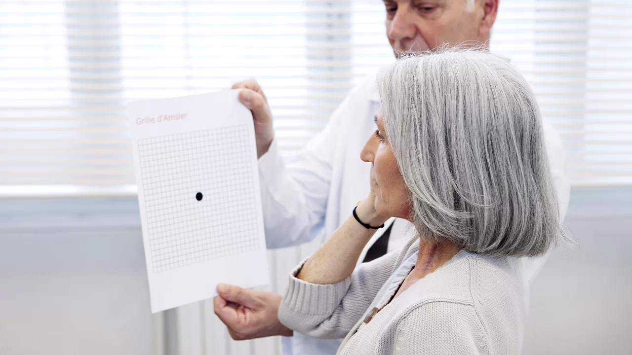
- Amsler Grid Test: With this simple test resembling a grid of squares, the patient's perception of straight lines is evaluated.
What are the Treatment Methods for Macular Degeneration?
The treatment plan is determined individually by Specialist Dr. Ayşe Öner based on the type of disease (dry or wet) and its stage of progression.
Dry Type Macular Degeneration Treatment:
Currently, there is no medical treatment or surgery that provides a complete cure for the dry type. However, to slow the progression of the disease and prevent it from turning into the wet type, special vitamin and antioxidant supplements compliant with the AREDS2 formula are prescribed.
Wet Type Macular Degeneration Treatment:
Treatment in the wet type aims to stop abnormal vessel development and prevent leaks.
- Intravitreal Injection Therapy (Anti-VEGF Therapy): This is the gold standard treatment. Special drugs are injected directly into the eye using very fine needles.
- Photodynamic Therapy (Cold Laser): In very specific cases, a drug injected into the eye is activated by a low-energy laser beam.
We are racing against time to protect your eyes and your quality of vision. If you have been diagnosed with macular degeneration or carry risks, you can contact us for a comprehensive evaluation and personalized treatment planning.
Frequently asked questions
Macular degeneration does not result in total darkness (total blindness); peripheral vision is always preserved. However, it destroys "central and detailed" vision required for reading, recognizing faces, and driving. In the "Wet Type" form, hemorrhaging is stopped and vision is preserved through new generation intravitreal Anti-VEGF (smart molecule) injections. In the "Dry Type" form, cellular death is slowed down and the patient's quality of vision is improved using innovative methods successfully applied in our clinic, such as Valeda Light Delivery System (Photobiomodulation).
Genetic predisposition is one of the major risk factors; however, it does not mean the disease will definitely manifest. The most critical step is detecting cellular stress before you experience any vision loss. With advanced technologies such as OCT (Optical Coherence Tomography/Optical Biopsy) and FAF (Fundus Autofluorescence/Cellular Metabolism Mapping) used in our clinic, we can detect the "footprints" of the disease years in advance and take immediate preventive measures.
We offer a "VIP Healing Package" with zero waiting time for our medical tourism patients. On the day of your arrival in Turkey, non-mydriatic (dropless) OCT and FAF scans are performed. If wet type is detected, a painless injection is administered within seconds on the same day, and you can safely board your flight. If dry type is detected, luxury hotel accommodations and VIP transfers for sessions of treatments like Valeda are organized by our assistants. Your treatment is completed in the comfort of a healing holiday.