The eye is like a deep ocean; it is not just the colored part we see from the outside, but a complex system housing a vast neurological network and delicate anatomy. Within these depths, issues occurring in the retina (the nerve layer)—such as tears, hemorrhages, or cellular tractions—are emergencies that carry a permanent risk of blindness and cannot be solved with drops or lasers alone. Vitrectomy (Vitreoretinal Surgery) is the pinnacle of eye surgery, requiring advanced specialization. It is a microsurgical miracle that allows us to reach these inaccessible depths and repair damage directly at the cellular level.
As Ophthalmology Specialist Dr. Ayşe Öner and her visionary clinical team—with international experience across thousands of vitreoretinal cases—we successfully treat the most severe conditions in our JCI-accredited, high-level operating theaters. By blending Turkey’s robust health tourism infrastructure with VIP comfort, we offer international patients immediate intervention, bypassing month-long waiting lists in their home countries for a flawless, stress-free healing journey.
What is Vitrectomy? A Journey to the Depths of the Eye
The interior of the eyeball is not empty; it is filled with a transparent, egg-white-like gel called the Vitreous. This gel is firmly attached to the retina at the back of the eye. Due to aging, trauma, or diabetes, this gel can lose its structure, fill with blood, or shrink and pull on the retina, causing a tear.
In its simplest definition, Vitrectomy is the microsurgical removal of this diseased or bloody vitreous gel, followed by the repair of the underlying retinal damage (tears, detachment, or membrane formation). When the gel is removed, the eye does not collapse; the body (or the surgeon) immediately fills this space with transparent intraocular fluids or special "tamponade" materials.
Targeted Conditions
Vitrectomy is the "heavyweight" surgery of ophthalmology, applied to the most serious threats to the posterior segment:
- Advanced Diabetic Retinopathy & Hemorrhages: Clearing dense internal bleeding (Vitreous Hemorrhage) caused by long-term diabetes. Leaky vessels are sealed with lasers during the same session.
- Retinal Detachment: A critical emergency where fluid leaks behind a retinal tear, peeling the nerve layer away from the eye wall. Vitrectomy allows us to reattach the retina to its original position.
- Macular Diseases: Closing holes in the visual center (Macular Hole) or peeling away wrinkling membranes (Epiretinal Membrane).
- Intraocular Foreign Bodies: Removing metal or glass shards from work accidents and reconstructing the shattered eye anatomy.
- Endophthalmitis (Infections): Rapidly cleaning infected gel and washing the interior with antibiotics.
- Cataract Complications: Safely retrieving lens fragments that have fallen to the back of the eye during unsuccessful cataract surgeries.
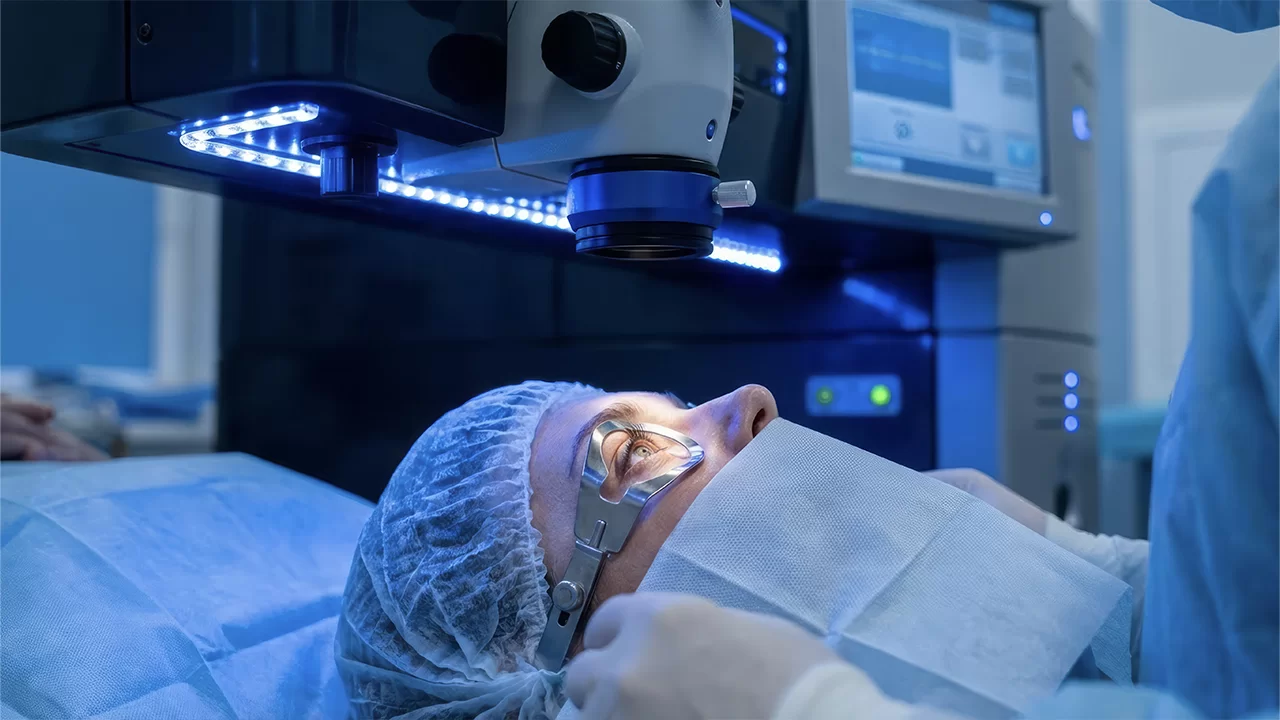
The Surgical Process: Sutureless Microsurgery
Modern technology has transformed vitrectomy into a sutureless, patient-friendly procedure:
- Anesthesia: Depending on the case, it is performed under local anesthesia (numbing around the eye) or general anesthesia.
- Micro-Incision (Pars Plana Vitrectomy): Three microscopic tunnels (23, 25, or 27 Gauge), thinner than a strand of hair, are opened in the white of the eye (sclera). These serve as channels for fluid, light, and surgical tools.
- Repair: Using high-magnification microscopes, the surgeon clears the gel, "solders" tears with a laser, or peels membranes.
- Tamponade (Gas or Silicone): To ensure the retina stays in place, the eye is filled with either special expanding gas or silicone oil. The incisions are so small that they usually close on their own without the need for stitches.
Post-Operative Detail: Positioning and Flight Warning
Success depends on the surgeon's skill and the patient's compliance:
- Positioning: If gas or silicone is used, the patient may need to maintain a specific position (usually face-down) for 1 to 2 weeks to ensure the retina reattaches correctly.
- Flight Warning: Patients with Gas in their eye strictly cannot fly or visit high-altitude mountains until the gas is fully absorbed (approx. 2–6 weeks). Pressure changes can cause the gas to expand, leading to a dangerous crisis. This restriction does not apply to silicone oil.
The VIP Advantage with Dr. Ayşe Öner
For an international patient with retinal detachment, waiting weeks for surgery can mean permanent blindness. Our clinic provides a high-stakes "Surgical Bridge":
- Zero Waiting, Life-Saving Urgency: When you contact us, your case is treated as a "Red Code." Your operating theater is reserved the moment you book your flight to Turkey.
- Expert Hands: Dr. Ayşe Öner’s vast experience ensures maximum success even in the most challenging detachment cases, with minimal tissue damage.
- International Logistics: If a gas tamponade is required, our health tourism assistants replan your stay. Since flying is prohibited, we organize luxury hotel stays for you and your companion, turning the mandatory waiting period into a comfortable recovery vacation.
- Native Language Support: Post-operative rules regarding "face-down" positioning and medications are explained clearly in your native language by our medical assistants, removing all anxiety.
Do not accept retinal damage as fate. To stop the path to blindness with the most advanced microsurgery and experience a VIP process with Turkey’s premier health tourism assurance, contact Specialist Dr. Ayşe Öner Clinic immediately. Vision is a right that often requires racing against time, and we are ready to win that race for you.
Frequently asked questions
Vitrectomy is the most advanced microsurgical method in ophthalmology. It involves removing the gel-like fluid (vitreous) from the eye to intervene in hemorrhages and tears at the back of the eye. Unlike traditional methods, this surgery is performed at the Dr. Ayşe Öner Clinic using the Sutureless (Stitchless) Vitrectomy technique. The procedure is completed through microscopic entries into the white of the eye, which are as thin as a strand of hair; these entries close on their own upon completion without the need for sutures. Consequently, post-operative pain, stinging, and redness are minimized.
When the retina (nerve layer) is damaged due to retinal tears, detachment, or severe diabetic hemorrhaging, it requires internal "tamponade pressure" to reattach to the eye wall and heal. Special expansive gases or silicone oils serve this purpose. When a gas tamponade is used, the body naturally absorbs and eliminates this gas within a few weeks. If silicone is used, a short secondary surgical procedure is required months later to remove the silicone once the retina's healing process is complete. The choice of tamponade is determined by your surgeon based on the specific condition of your ocular damage.
In vitrectomy surgeries, if "gas" is placed in your eye, flying is strictly prohibited due to cabin pressure risks until the gas is completely absorbed (usually between 2 to 6 weeks). Our VIP medical tourism ecosystem resolves this without letting it turn into a crisis. Our assistants organize a comfortable convalescence period for you and your family in a luxury hotel until your flight ban ends. If "silicone" is placed in your eye, there is no contraindication to flying as silicone is not affected by pressure, and you can safely return to your country a few days after surgery.