Our eyes, which allow us to perceive the world with all its colors, depth, and sharpness, possess an extremely delicate and flawless balance at a microscopic level. However, advancing age and various environmental factors can lead to undesired structural changes in this sensitive balance. The Epiretinal Membrane, which forms at the very center of vision and distorts visual quality as if looking through wrinkled glass, is a condition that significantly reduces patients' quality of life but can be successfully treated with modern microsurgical methods.
What is Epiretinal Membrane (ERM)?
Also known in medical literature as "Macular Pucker," "Cellophane Maculopathy," or "Premacular Fibroplasia," Epiretinal Membrane (ERM) is a thin, semi-transparent, and membrane-like scar tissue that forms directly over the retina—the innermost layer of the eye—and specifically the macula (yellow spot) region responsible for sharp, detailed vision.
To better understand this condition, you can imagine a thin, transparent piece of cellophane tape sticking onto the retina, which we compare to the film of a camera. Initially just a transparent membrane, this tissue begins to shrink and contract over time. This shrinking membrane mechanically pulls and wrinkles the delicate macular tissue beneath it, disrupting the smooth anatomical structure of the retina. This physical wrinkling and traction on the retina cause significant fluctuations, blurriness, and distortions in the patient's central vision.
What are the Causes and Risk Factors?
The development of an epiretinal membrane is mostly a natural consequence of the aging process. In a large majority of patients, there is no specific underlying disease triggering the membrane formation; this condition is called "Idiopathic Epiretinal Membrane." The primary factors and risk factors in the formation mechanism are:
- Posterior Vitreous Detachment (PVD): The jelly-like fluid called vitreous liquefies and shrinks with age. As it separates from the retina, it can sometimes cause microscopic damage or cell shedding. The eye sends reparative cells to the area as a defense mechanism, which can grow out of control and form an undesired scar tissue (membrane) on the macula.
- Prior Eye Surgeries: The risk of developing an epiretinal membrane increases as a healing response following surgeries such as cataract surgery, retinal detachment surgery, or laser interventions.
- Intraocular Inflammation (Uveitis): Chronic inflammation in the inner layers of the eye creates a basis for cell accumulation and membrane formation.
- Retinal Vascular Diseases: Diseases that cause bleeding and leakage, such as diabetic retinopathy or retinal vascular occlusions, are factors that strongly trigger membrane formation.
- Eye Trauma: Severe blunt blows to the eye can lead to the formation of traumatic healing tissue on the retinal surface.
What are the Symptoms of Epiretinal Membrane?
The disease usually progresses slowly and insidiously. In the early stages, when the membrane is still thin and has not yet shrunken, the patient may feel no complaints. However, as the membrane thickens and begins to wrinkle the retina, the following specific symptoms appear:
- Distorted and Curved Vision (Metamorphopsia): The most typical complaint. Straight lines (e.g., window edges, door frames, or tile lines) appear curved, wavy, or bent.
- Blurred Central Vision: A decrease in the clarity of the focus point, creating a foggy or hazy visual field.
- Macropsia or Micropsiva: Perceiving objects as larger or smaller than their actual size.
- Double Vision (Monocular Diplopi): Seeing objects as double or shadowed even when only one eye is open.
- Difficulty with Fine Details: Significant difficulty in threading a needle, reading small print, or discerning fine details on human faces.
High-Tech Diagnostic Methods
The diagnosis of epiretinal membrane and surgical planning are carried out with high-tech devices requiring micron-level precision:
- Detailed Fundus Examination: The pupil is dilated with drops, and the specialist directly detects the cellophane-like membrane and the wrinkles it creates using special lenses.
- Optical Coherence Tomography (OCT): This is the most critical, radiation-free 3D imaging technology for diagnosis and follow-up. The thickness of the membrane, how much it pulls the retina, and whether it creates macular edema (fluid accumulation) are mapped at a cellular level. Surgical decisions are largely based on OCT results.
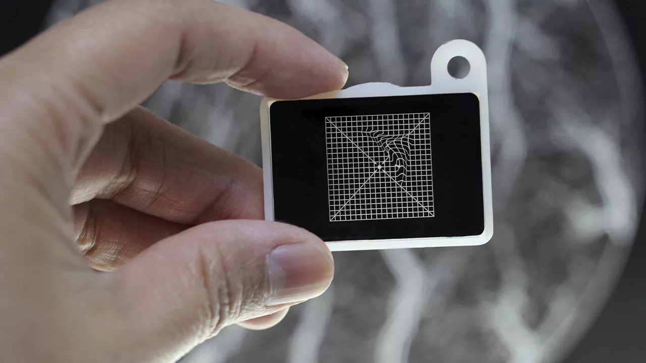
- Amsler Grid Test: A standard grid paper test used to measure the degree of curved and wavy vision.
Treatment: Microscopic Surgery
It is not possible to treat an epiretinal membrane with medication, eye drops, glasses, or laser. The method and timing of treatment are determined by the severity of the patient's complaints and the damage the membrane has caused to the retina.
Observation and Follow-up
If the membrane is very thin, does not affect the patient's daily life, and has not created serious traction on the retina, surgery is not immediately decided upon. The progression of the membrane is monitored with regular OCT scans.
Pars Plana Vitrectomy and Membrane Peeling Surgery
If the membrane has thickened, significantly reduced vision quality, and created a pucker on the retina, the only treatment is surgery. This operation is one of the most delicate and skill-intensive procedures in eye surgery:
- Through millimetric, sutureless incisions in the white part of the eye (sclera), the vitreous gel is completely removed.
- The surgeon reaches the retina using special microscopes and intraocular lighting systems.
- Using special micro-forceps thinner than a human hair, this pathological membrane is "peeled" from the retina with extremely slow and gentle movements.
- Once the membrane is peeled, the mechanical pressure on the retina is eliminated, and the tissue gradually returns to its natural, smooth anatomical shape.
Visual quality and distorted vision complaints improve gradually over weeks and months following the surgery.
Do not let distortions in your vision overshadow the truths of your life. To entrust your eyes to the most advanced technology and reliable surgical hands, contact our patient department.
Frequently asked questions
Yes, this "distorted vision" (Metamorphopsia) is the most typical and primary symptom of Epiretinal Membrane (macular pucker). A transparent membrane left by the vitreous gel on the macula contracts over time, wrinkling the underlying retinal layer like a piece of paper. This anatomical wrinkling distorts your vision like a funhouse mirror. A definitive diagnosis is made with an OCT scan lasting seconds.
This operation is the most delicate micro-surgery in ophthalmology, requiring "jewelry-grade craftsmanship" (Vitrectomy with Membrane Peeling). In the hands of the right surgeon, it is not dangerous; on the contrary, it is a highly safe micro-surgery with satisfying results. Dr. Ayşe Öner peels this wrinkled membrane perfectly using special biological dyes and microscopic forceps without causing even micron-level damage to your vision cells.
As soon as the surgery is finished, the membrane wrinkling your retina is physically removed (anatomical success). However, it takes weeks, sometimes months, for the wrinkled retinal tissue to flatten out like a bedsheet and for the brain to adapt to this new flat image. The healing process is gradual. Our international patients can follow this process remotely and safely via OCT results sent through our Tele-medicine system.