Our eye health is the fundamental key to maintaining an independent and high-quality daily life. Even the slightest damage occurring in our central visual field, which allows us to see clearly the point we focus on, can make many activities—from reading to driving—impossible. A Macular Hole, which can emerge with advancing age or various eye traumas and directly threatens central vision, is a condition that can be treated with timely and accurate surgical intervention.
What is a Macular Hole?
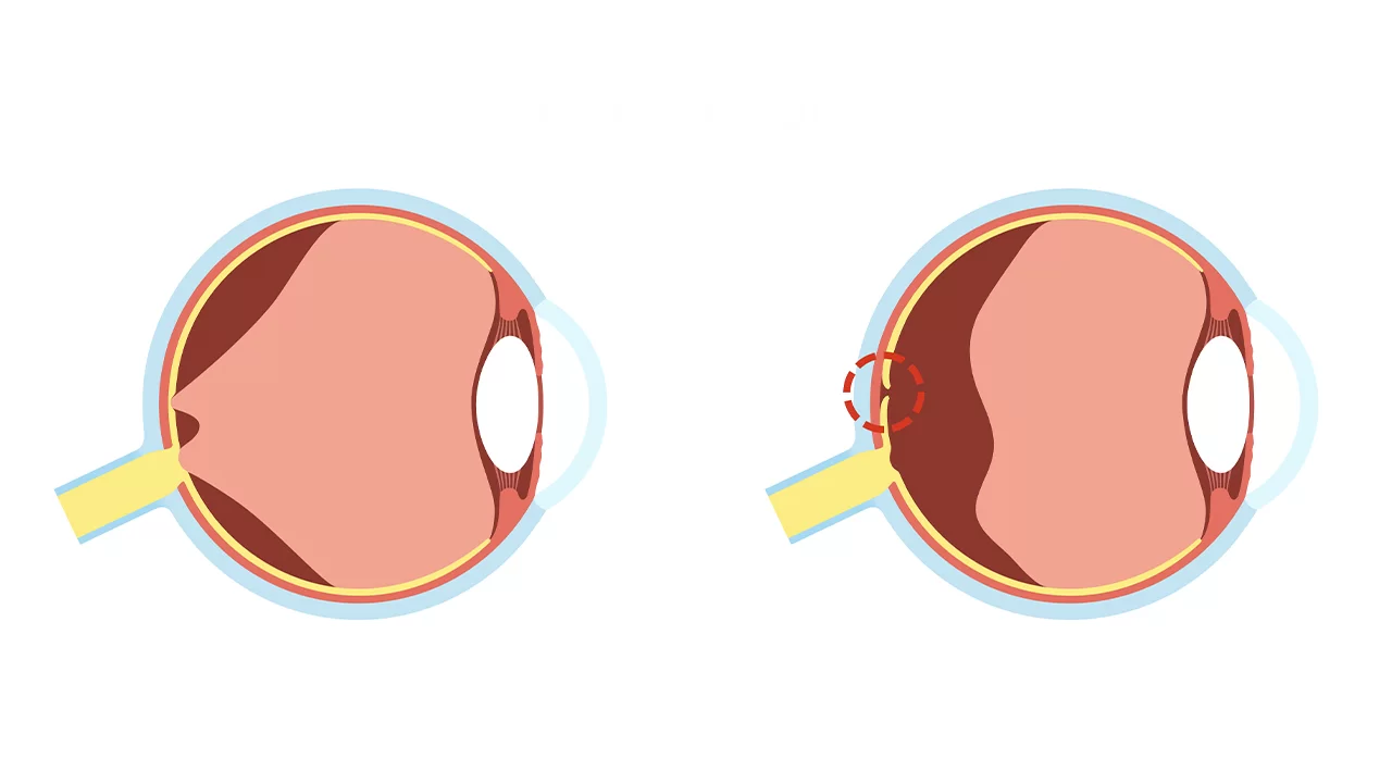
The nerve layer covering the back inner wall of our eye, consisting of light-sensitive nerve cells, is called the retina. The millimetric area located at the very center of the retina, approximately the size of a pinhead, is called the macula (yellow spot). The macula is the most vital region responsible for "central vision," providing our highest visual acuity and allowing us to recognize fine details, colors, and faces.
A Macular Hole, as the name suggests, is the formation of an anatomical tear—a "hole"—caused by the separation of nerve cells at the very center of this sensitive and critical region. Due to this hole, the photoreceptor cells in the macula lose their integrity, and images at the patient's point of focus are transmitted to the brain as incomplete or distorted. Consequently, while peripheral vision remains intact, a serious loss, blurriness, or a dark spot appears in the central vision.
What are the Causes and Risk Factors?
The most fundamental factor in the development of a macular hole is the aging process of the eye. The disease usually appears in individuals aged 60 and over and is slightly more common in women. The formation mechanism is based on these primary causes:
Vitreomacular Traction (VMT)
The jelly-like structure that fills most of the eye and maintains its spherical shape is called the vitreous gel. In younger years, this gel is firmly attached to the retina. As we age, it undergoes structural changes, liquefying and shrinking. The shrinking vitreous slowly separates from the retina (Posterior Vitreous Detachment). However, in some cases, this gel adheres to the macula much more firmly than normal. During the separation process, it pulls the macula outward like a suction cup. Over time, this traction force causes the macular tissue to tear and a hole to open in its center.
Other rarer risk factors include:
- High Myopia: An eyeball longer than normal leads to stretching and thinning of the retina, creating a basis for hole formation.
- Eye Trauma: Severe blunt blows to the eye (e.g., ball impact or traffic accidents) can cause sudden macular tears.
- Prior Eye Surgeries: The risk increases in eyes with a history of complicated cataract surgery or retinal detachment.
- Macular Edema: Long-term macular swelling (edema) can trigger hole formation by disrupting the tissue structure.
What are the Symptoms of a Macular Hole?
The formation of a macular hole is a completely painless process. Since the disease usually starts in one eye and the other eye is healthy, patients may not notice the condition in the early stages. Symptoms include:
- Blurred Central Vision: Loss of clarity at the point of focus, feeling like looking through foggy glass.
- Distorted Vision (Metamorphopsia): Straight lines, such as door frames or tile lines, appearing wavy, crooked, or bent.
- Difficulty Reading: Letters skipping, merging, or becoming unreadable while reading or using a phone.
- Central Dark Spot (Scotoma): As the hole grows, a fixed gray or black spot appears in the center of the visual field. When looking at a person's face, the patient cannot see the face itself, only the shoulders and surroundings.
Our Advanced Diagnosis Methods
In eye health, early diagnosis is the foundation of surgical success. In our clinic, we use the most up-to-date diagnostic tools:
- Dilated Fundus Examination: The macula is directly examined with special lenses after dilating the pupils.
- Optical Coherence Tomography (OCT): This is the gold standard for diagnosing a macular hole. It provides microscopic, 3D cross-sections of the retina in seconds without radiation. The hole’s diameter, depth, and stage are measured with micrometer precision. Surgical decisions are made entirely based on this high-resolution mapping.
- Amsler Grid Test: Used to measure the degree of distortion and fluctuations in the patient's central vision.
Treatment Methods: Surgical Excellence
It is not possible to treat a macular hole with medication, drops, or glasses. The only definitive solution is the microsurgical method requiring advanced technology.
Pars Plana Vitrectomy Surgery:
Macular surgery is one of the most delicate operations in ophthalmology. Dr. Ayşe Öner’s experience with thousands of vitrectomy cases carries our success rates above world standards. The process involves:
- Vitreous Removal: Entering the eye through sutureless incisions (approx. 0.4 mm). The vitreous gel causing the traction is completely removed.
- ILM Peeling: To ensure the hole closes, an extremely thin layer on the top of the retina (Internal Limiting Membrane - ILM) is gently peeled using micro-forceps. This is the most critical and sensitive stage of the surgery.
- Gas Tamponade: A special medical gas is filled into the eye to act as an internal "plug" (tamponade), pushing the edges of the hole together to allow them to heal.
Importance of the "Face-Down Position"
For the gas to press against the macular hole at the back of the eye, the patient must utilize gravity. Therefore, it is vital for patients to spend most of the day (including sleep) in a face-down position for 3 to 7 days post-surgery. The gas is absorbed by the body over weeks and replaced by the eye's natural fluid.
Do not let damage in your vision center settle in the center of your life. Contact our patient coordinators immediately to regain your health with the world's best technology and experienced hands.
Frequently asked questions
A macular hole, which is an anatomical tissue disruption in the yellow spot (macula), cannot be treated with drops, medication, or lasers. The probability of spontaneous closure (except for very early initial stages) is near zero. The only definitive treatment in the world is the micro-surgical method known as Vitrectomy and Membrane Peeling.
During surgery, after the microscopic membranes around the hole are cleared, an expansive gas is filled into the eye. This gas bubble acts as a tamponade, applying internal pressure to the open hole and allowing the tissues to adhere and close. Because the gas is light and needs to apply pressure upward (toward the back of the eye), it is vital to lie face-down (looking at the floor) for usually 5 to 7 days after surgery. This is the most critical factor determining the success of the surgery.
Your surgery is performed by expert hands with zero waiting time. However, flying is prohibited because there is gas in your eye. Our assistants organize a "convalescence stay" for you in a luxury hotel near our clinic. You complete the face-down positioning process in the comfort of a home. Once the flight ban period ends or via alternative safe land/sea transfers, your return to your country is planned step-by-step by our medical tourism team.