The optic nerve (visual nerve), which provides perfect visual communication between our eyes and the outside world and functions as the body's most complex technological data cable, is the fundamental structure that builds our visual world. However, when this vital transmission line is damaged for various reasons, serious losses in vision quality—which can develop suddenly or slowly and sometimes be permanent—may occur. This group of diseases, gathered under the umbrella of Optic Neuropathies in medical literature, requires neuro-ophthalmology expertise and a multidisciplinary, comprehensive approach.
What is Optic Neuropathy?
The optic nerve is a massive network of approximately 1.2 million fine nerve fibers that collects light and color signals formed in the retina (the back layer of the eye) and transmits them to the visual center (occipital cortex) at the back of the brain. It is not enough for the eye to record the image like a camera; for this image to be interpreted, it must reach the brain completely via the optic nerve.
The word "neuropathy" refers to a nerve becoming diseased, losing its function, or being damaged by external factors. Therefore, Optic Neuropathy is not the name of a single disease but a general umbrella term for a wide range of conditions that cause cellular damage and disconnection in the visual nerve. When nerve fibers become inflamed, deprived of oxygen, or die due to injury (optic atrophy), visual signals to the brain are interrupted, resulting in serious visual impairments ranging from blurred vision to total blindness.
Types and Causes of Optic Neuropathy
The mechanism of optic nerve damage varies greatly from patient to patient. Identifying the specific type is vital for an accurate treatment plan. The most common types managed at our clinic include:
1. Ischemic Optic Neuropathy
This is damage caused by the sudden interruption or reduction of blood flow in the microscopic capillaries nourishing the optic nerve (similar to a heart attack or stroke), leading to oxygen deprivation. It is usually seen in individuals over 50. Risk factors include hypertension, diabetes, high cholesterol, atherosclerosis, sleep apnea, and smoking. Patients usually notice painless, sudden vision loss in one eye upon waking up.
2. Optic Neuritis (Inflammatory Optic Neuropathy)
This is an inflammation caused by the immune system mistakenly attacking its own healthy nerve cells. It is frequently seen in young adults (ages 20-40) and more commonly in women. It may appear as the first sign of Multiple Sclerosis (MS) or develop due to conditions like Neuromyelitis Optica (NMO) or viral infections. The most typical feature is severe pain that increases with eye movements and blurred vision.
3. Compressive Optic Neuropathy
This occurs when the optic nerve is mechanically crushed or subjected to physical pressure by a mass along its anatomical path to the brain. Brain or orbital tumors, cysts, aneurysms, or the swelling of eye muscles due to Thyroid Eye Disease (Graves) can cause slow and stealthy vision loss.
4. Traumatic Optic Neuropathy
This involves the physical tearing, excessive stretching, or compression of the optic nerve due to traffic accidents, falls, or severe blunt and penetrating injuries to the head or eye. It is a severe condition requiring emergency intervention with a high risk of blindness.
5. Hereditary Optic Neuropathies
These occur when optic nerve cells are born with structural defects or lose function at an early age due to genetic mutations. Leber’s Hereditary Optic Neuropathy (LHON) and Dominant Optic Atrophy are the most well-known examples, often causing painless and progressive central vision loss in both eyes symmetrically.
Symptoms of Optic Neuropathies
Symptoms can develop suddenly (within hours) or slowly (over months/years) depending on the type. Characteristic signs include:
- Decreased Visual Acuity: Levels ranging from slight haziness to perceiving only light or total darkness.
- Color Vision Impairment (Dyschromatopsia): Particularly perceiving red and green tones as faded or grayed out (Red desaturation). This is one of the earliest signs.
- Pain During Eye Movements: Especially in Optic Neuritis, patients feel a deep, pulling pain behind the eye when looking in different directions.
- Visual Field Losses: A fixed dark spot in the center (central scotoma) or a horizontal darkening in the upper or lower half of the vision.
- Impaired Pupil Reflex: The pupil failing to constrict properly or paradoxically dilating when light is shone into the affected eye (Relative Afferent Pupillary Defect - RAPD).
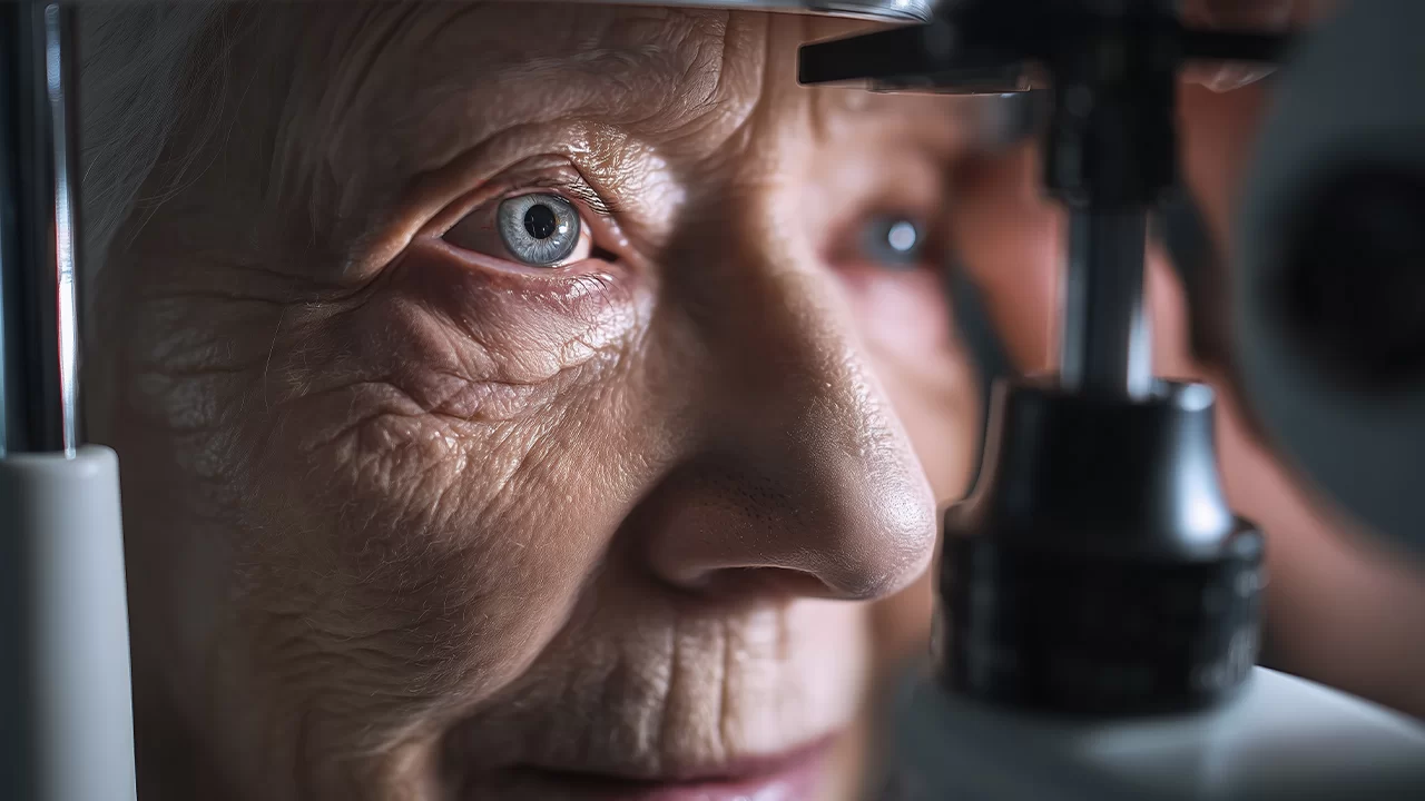
Advanced Diagnosis and Neuro-Ophthalmological Analysis
Since optic neuropathies can be easily confused with other retinal or central nervous system issues, a specialized Neuro-Ophthalmology unit is required. At Dr. Ayşe Öner’s clinic, we analyze these conditions at cellular and electrical levels:
- Fundus Examination: Direct inspection of edema (swelling), hemorrhage, or pallor (atrophy) of the optic disc.
- Optical Coherence Tomography (OCT): 3D measurement of the Retinal Nerve Fiber Layer (RNFL) thickness with micron precision to objectively show nerve thinning or edema.
- Visual Evoked Potentials (VEP): Measuring the speed and neural strength of electrical conduction. This definitively detects inflammation (demyelination) or damage.
- Computerized Visual Field (Perimetry): Mapping microscopic blind spots that the patient may not yet have noticed.
- MRI and Blood Tests: Brain and orbital MRIs are essential to rule out MS plaques, aneurysms, or tumors. Genetic tests and B12 levels are also meticulously investigated.
Treatment Methods and Multidisciplinary Approach
The primary goal of treatment is to protect surviving nerve tissue, resolve acute edema, and prevent progression.
- Inflammatory Neuropathies: The gold standard is high-dose Intravenous Steroid (Pulse Cortisone) treatment to rapidly resolve aggressive inflammation.
- Ischemic Neuropathies: Controlling systemic risk factors is essential. Blood thinners and vascular protective treatments are planned in coordination with neurology and cardiology units.
- Compressive Neuropathies: If a mass is physically crushing the nerve, emergency microsurgical decompression is performed in collaboration with neurosurgery or oculoplastic specialists.
- Visual Rehabilitation: For patients with permanent loss, the process is supported by customized telescopic systems, magnifying lenses, and contrast-enhancing filters to promote independent living.
Contact our expert assistants immediately to illuminate the shadows on your optic nerves with the power of science and experience a world-class, stress-free journey.
Frequently asked questions
Your eye may be a perfect camera, but if the USB cable that transmits the photo taken by that camera to the computer (the brain) is disconnected or damaged, the screen remains dark. Optic neuropathy is the damage to that main transmission cable (the optic nerve). Conditions such as Multiple Sclerosis (MS), Optic Neuritis (nerve inflammation), past accidents/traumas, genetic mutations (LHON), or pressure from brain tumors cause this damage, typically creating foggy/dark vision in the center and fading of colors.
Pallor (atrophy) in the optic nerve does not necessarily mean that 100% of the fibers within the nerve are dead. What matters is the detection of "remaining viable fibers." With VEP (Electrophysiological Mapping) performed in our clinic, the electrical conduction capacity of the nerve is objectively measured. If "living cells" are still detected in the nerve, advanced Magnovision treatments, which strengthen and retrain that electrical line between the brain and the eye on a cellular basis, are initiated to start the rehabilitation process.
Absolutely not. The Dr. Ayşe Öner Clinic is a sub-specialty center that complements your systemic neurological treatment and focuses entirely on your optic nerve. When you contact our medical tourism team, your VIP airport greeting and luxury hotel stays, which reduce stress factors to zero, are pre-planned. In our clinic, your nerve map is created with the help of medical translators who speak your native language fluently, and your session-based treatment (neuro-rehabilitation) protocols to re-awaken that damaged cable begin immediately in VIP comfort.