Our eyes are home to one of the most complex and perfectly functioning microscopic vascular networks in our body. Even the slightest disruption in this delicate vascular system can directly and sometimes irreversibly affect vision quality. Retinal Vascular Occlusions, which can also be described in medical literature as a type of "eye stroke," are among the most common causes of sudden vision loss.
What is Retinal Vascular Occlusion?
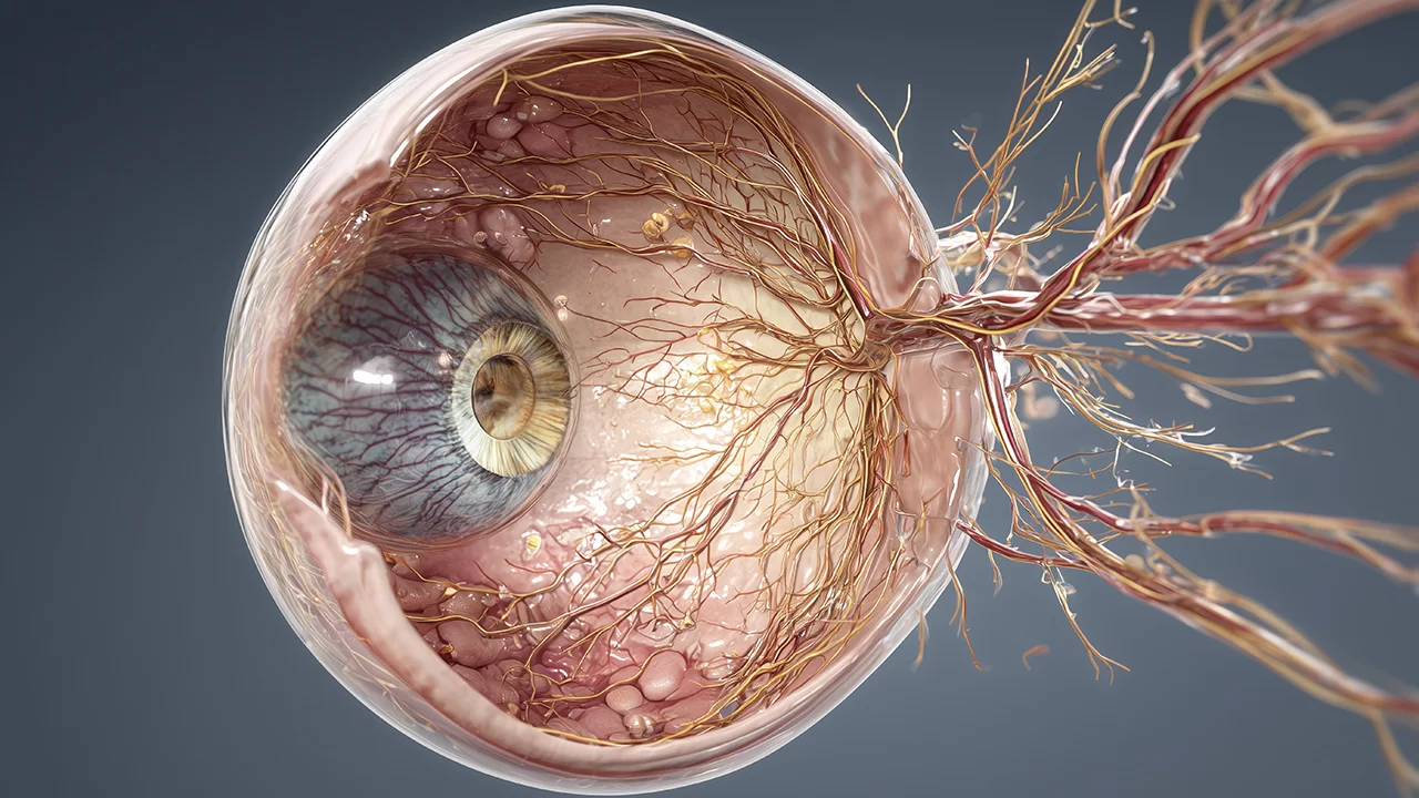
Just as our heart or brain needs an uninterrupted blood flow to function, the nerve layer of our eye (retina) similarly requires rich oxygen and nutrient support. The retina is nourished by a main artery (artery) coming from the center and sends back used, deoxygenated blood through a main vein (vein) also exiting from the center. These main vessels divide into smaller branches as they spread across the retinal surface.
Retinal vascular occlusion is a condition where one of the arteries or veins of the retina becomes blocked due to a clot, calcification, or external pressure, much like a heart attack or a stroke in the brain. As a result of the blockage, the retinal tissue either cannot receive the oxygen it needs or, because it cannot remove the dirty blood, blood leaks into the eye and edema (swelling) forms in the tissues.
What are the Types of the Disease?
The disease is divided into four main categories based on the type of vessel blocked (artery or vein) and its location (main vessel or branch), and the clinical picture of each is different:
1. Central Retinal Artery Occlusion (CRAO)
This is one of the most urgent emergencies in eye health. It is the sudden blockage of the main artery that brings fresh blood and oxygen to the eye, usually by a clot (embolism) originating from the neck or heart. The retina is deprived of oxygen within seconds. Without feeling any pain, the patient experiences total or near-total vision loss in that eye within seconds. Since irreversible damage starts very quickly, intervention within the first few hours is vital.
2. Branch Retinal Artery Occlusion (BRAO)
This is the blockage of one of the smaller branches of the main artery spreading to the retina. Cell death occurs in the retinal region nourished by the blocked vessel. The patient does not experience total blindness but feels a sudden darkening or a curtain falling over a part of the visual field.
3. Central Retinal Vein Occlusion (CRVO)
This is the blockage of the main vein that removes dirty blood from the vision center. Since blood cannot exit the eye, pressure inside the vessel increases, vessels rupture, and widespread bleeding and fluid leakage into the retinal tissue begin. This situation leads to severe swelling in the macula (our sharp vision point), known as Macular Edema.
4. Branch Retinal Vein Occlusion (BRVO)
This is the blockage of only one branch of the vein. Bleeding and fluid leakage are limited only to the retinal area where the blockage is located. It is the most common type of vascular occlusion. If the fluid leakage does not reach the macula, the patient may not notice the condition.
What are the Causes and Risk Factors?
Retinal vascular occlusions usually appear as a reflection of systemic vascular diseases in the body rather than the eye itself. The most prominent risk factors are:
- Hypertension (High Blood Pressure): The factor that most increases the risk by causing hardening and narrowing of the vessel walls.
- Atherosclerosis and High Cholesterol: Plaque formations within the vessels obstruct blood flow.
- Diabetes: Destroys elasticity by damaging the vascular structure at a cellular level.
- Heart Diseases: Heart valve diseases or rhythm disturbances are the primary causes of clots traveling to the eye.
- Glaucoma (Eye Pressure): High internal pressure slows blood flow by applying mechanical pressure to the vessels in the optic nerve.
- Smoking and Advanced Age: Universal risk factors that directly threaten vascular health.
What are the Symptoms of the Disease?
The most characteristic feature of retinal vascular occlusions is that they are painless. Symptoms vary depending on the type of blockage:
- Sudden total vision loss in one eye.
- Dark areas or shadowing in part of the visual field.
- Gradually increasing or suddenly developing blurred or hazy vision.
- Straight lines appearing crooked, bent, or wavy.
- Floating black spots or spider webs in advanced vein occlusions.
Our Advanced Diagnosis Process
To determine the treatment strategy, it is necessary to fully map the extent of the damage. Our comprehensive diagnostic methods include:
- Dilated Fundus Examination: Retinal hemorrhages, "cotton-wool" spots, and vascular tortuosity are directly detected.
- Fluorescein Angiography (FFA): The gold standard in determining the type and location of the blockage. Ischemic (non-perfused) areas and leaking vessels are visualized in detail.
- Optical Coherence Tomography (OCT): Measures the fluid (macular edema) accumulated in the yellow spot at the micron level.
- Systemic Evaluation: Our patients are consulted with Cardiology and Internal Medicine specialists to coordinate the treatment of the underlying systemic disease.
Retinal Vascular Occlusion Treatment Methods
The main goal of treatment is to dry the macular edema and prevent complications.
Vein Occlusion Treatment:
- Intraocular Injections (Anti-VEGF and Steroids): The most effective method to treat leakage and macular edema. These injections, performed with very fine needles, rapidly reduce fluid accumulation.
- Argon Laser Photocoagulation: If ischemic areas are detected, laser is applied to these regions to prevent the formation of new abnormal vessels that could lead to blindness or painful glaucoma.
Artery Occlusion Treatment:
Central artery occlusion is a race against seconds. Emergency medical interventions such as lowering intraocular pressure and massaging the eyeball to push the clot to more distant branches are applied within the first 24 hours.
If you feel a sudden loss of vision or blurriness, do not take it lightly. You can contact our health assistants immediately to take the most accurate and fastest step toward protecting your vision.
Frequently asked questions
Yes, it is usually a severe loss of vision that develops suddenly, often upon waking in the morning or during the day, without any pain. As a result of the blockage of the central or branch retinal veins, the interior of the eye suffers from extensive hemorrhage and edema. If intervention is made quickly within the first hours or days using intravitreal injections (Anti-VEGF) and lasers, the edema is resolved and vision can be largely salvaged.
Retinal vascular occlusions are actually a warning from the body (the systemic system), not just the eye. Hypertension, diabetes, high cholesterol, or cardiovascular diseases are the primary causes. To prevent the disease from occurring in the other eye or recurring, we do not just treat your eye; we ensure your systemic diseases are brought under control in coordination with internal medicine or cardiology departments.
Eye stroke is a race against time. The minute our international patients reach the clinic, the occluded vessel is mapped in seconds using our dye-free OCT-A (Optical Coherence Tomography Angiography) technology. Your intravitreal injection treatment is administered within the same hour to stop the bleeding. You achieve healing at VIP speed without losing time (and vision cells) in long equipment queues.